 |
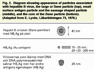
Subsequently Dane,
Cameron and Briggs identified a larger particle about 42 nm in diameter with
an electron dense core of about 27 nm (25). It is probable that this
represents the whole virus particle. Both the 20 nm and 42 nm particles
contain Australia antigen on their surfaces and this is now termed hepatitis
B surface antigen (HBsAg). The surface antigen can be removed from Dane
particles by the action of detergents to reveal the core which has its own
antigen, hepatitis B core antigen (HBcAg). Antibodies to both these antigens
(anti-HBs, anti-HBc) can be detected in human blood. The surface antigen can
be detected in the peripheral blood by the methods we initially introduced
and by more sensitive methods which have since been developed. Anti-Hbs is
often found in the peripheral blood after infection and may persist for many
years. It may also be detected in people who have not had clinical
hepatitis. Anti-HBc is usually associated with the carrier state (i.e.
persistent HBsAg in the blood), but may occur without it. HBcAg itself has
not been identified in the peripheral blood. Anti-HBc is also found commonly
during the active phase of acute hepatitis, before the development of anti-HBs
but in general does not persist as long as anti-Hbs. |