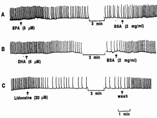
Figure 3. Effects of EPA and DHA on spontaneous contraction rates of
cultured neonatal rat cardiomyocytes. Perfusion of the myocytes with 5
µM EPA (A) or of DHA (B) slowed the
beating rate within 2 minutes. Addition of delipidated bovine serum albumin
(2 mg/ml) to superfusate extracted the EPA or DHA and returned contraction
rate to control levels, Tracing C shows similar effects of lidocaine 20
µM on spontaneous contraction rate. |
Fig. 3 shows the characteristic slowing of the
beating rate of the myocytes when low µM
concentrations of EPA or DHA were added to the medium bathing the isolated
heart cells. When delipidated bovine serum albumin, which has three high
affinity binding sites for fatty acids, was added to the superfusate, the
EPA or DHA was extracted from the heart cells and the beating rate returned
to the control rates. So the slowing effect of the fatty acids on the
beating rate is reversible. Toxic agents, known to produce fatal arrhythmias
in humans, were added to the medium bathing the cultured cells and the
effects of adding the n-3 fatty acids observed. So we tested increased
extracellular Ca2+ , the cardiac glycoside ouabain, isoproterenol,
lysophosphatidyl choline and acyl-carnitine, thromboxane, and even the Ca 2+
ionophore A23187. All of these agents induced tachyarrhythmias in the
isolated myoctes.
|
