| front |1 |2 |3 |4 |5 |6 |7 |8 |9 |10 |11 |12 |13 |14 |15 |16 |17 |18 |19 |20 |21 |22 |23 |24 |25 |26 |27 |review |
 |
NIH. Consensus Conference Statement. 2000;17(1):1-36. WHO.
Available at: http://www.who.int/hrp/progress/40/prog40.pdf.
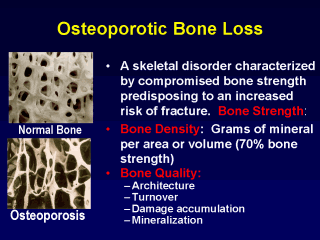
These figures illustrate the changes in bone architecture at the microscopic level that are characteristic of osteoporosis. Normal trabecular bone, as shown on the left, appears as a dense network of thick trabeculae with small spaces. Osteoporotic trabecular bone, on the right, shows clear loss of bone, with large spaces and perforation of the trabecular network (see arrows). These perforations lead to weakened bone, and ultimately to fractures. This bone loss and architectural damage is relatively rapid in the perimenopausal phase of PMO and has been reported to be rapid in patients initiating chronic glucocorticoid therapy. |